
In healthy individuals, as in dyslexics, the left asymmetry of a temporal area involved in the processing of language sounds is associated with better verbal performances. This finding was made possible by taking account of individual anatomical variability in a database with a large number of participants.
Since Geschwind in 1968, the localization in the left hemisphere of the language areas is associated with the anatomical lateralization of the planum temporale, a flat cortical area located on the upper surface of the temporal lobe and larger in the left hemisphere in 70% of cases. This hypothesis, deduced only from anatomical observations, is coherent with the lower leftward asymmetry of the planum temporale reported in dyslexic persons. But what about healthy people? For the first time, our study shows a link between the leftward asymmetry of the planum temporale and global verbal skills such as memory skills, phonological skills or the vocabulary extent…
Taking into account inter-individuals anatomical variability of the temporal lobe
Until now, it was not possible to explain the paradox raised by Bishop in 2013 on the lack of relationship between the leftward asymmetry of the planum temporale and the linguistic abilities in healthy persons. At the anatomical level, it is important to underline that the planum temporale neighboring the Heschl gyrus, a cerebral structure that contains the primary auditory area. Heschl’s gyrus is subjected to a very high anatomical variability: it can be single or double and its duplication may be total or partial. While some authors consider that in the case of duplication the second Heschl gyrus belongs to the planum temporale, others include it in the planum temporale definition only when the duplication is complete. The definition of the planum temporale varying from one study to another, it is difficult to find one’s way, and one understands that the way in which the various configurations of the Heschl gyrus are taken into account in the anatomical definition of the planum temporale directly impacts the size of the latter. To precisely measure the surface of the planum temporale and its asymmetry, an expert drew it on each of the anatomical images obtained by MRI in the 428 participants, carefully taking into account the number of duplications of Heschl’s gyrus. This led to 2 anatomical definitions of the planum temporale: 1) the posterior planum temporale alone, 2) the posterior planum temporale associated with the second Heschl’s gyrus. This way, it was possible to detect that individuals with an asymmetry of the posterior planum temporale alone (whether the asymmetry is rightward or leftward) have better verbal test performance than others.
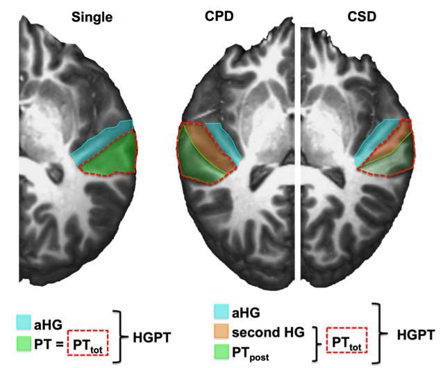
Illustration of the different definitions of the planum temporale depending on Heschl’s gyrus (HG) gyrification pattern. The knife-cut method was used to reveal the supratemporal plane, thanks to a slice passing through Heschl’s gyrus and through the ending of the Sylvian fissure. Three configurations are shown. The left panel shows a left hemisphere with a single HG colored in light blue and the planum temporale in green. On the middle panel, a complete posterior duplication of HG (CPD) is shown on the left (right hemisphere) with the first HG colored in light blue, the second HG in orange, and PTpost in green and a partial duplication (CSD) on the right (left hemisphere). A red dotted line delineates the whole PT (PTtot), corresponding to PT in case of single gyrus or including PTpost and the second gyrus in case of duplication. The HGPT area corresponds to the union of all colored areas including HG and the PT whatever the HG gyrification pattern.
Increase sensitivity by including a large population allowed to uncover the importance of asymmetry, whatever its side
Previous studies on these issues included at best a few dozen participants. Our study is based on a large sample of 428 healthy adults belonging to the BILGIN, a database specifically dedicated to the study of the anatomical and functional lateralization of the brain. To eliminate the related effects of sex or manual preference, the BILGIN includes as many men as women and as many right and left handed. The 428 participants each had an anatomical MRI and were assessed on their verbal skills. The association between the improvement of verbal performances and lateralization of the BILGIN is of low amplitude but very stable. It concerns all measured verbal competences: extent of vocabulary, verbal memory, phonological capacities … Without this large number of participants in the study, the asymmetry of the planum temporale as an element optimizing cerebral organization for language would have go unnoticed in healthy individuals. This is in accordance with previous results on language functional lateralization, rightward as leftward individuals performing better in cognitive tasks than individuals with low lateralization (Mellet 2014).
Bibliography
Bishop DVM (2013) Cerebral Asymmetry and Language Development: Cause, Correlate, or Consequence? Science 340:1230531.
Geschwind N, Levitsky W (1968) Human brain left-right asymmetries in temporal speech region. Science 161:186-187.
Marie D, Jobard G, Crivello F, Perchey G, Petit L, Mellet E, Joliot M, Zago L, Mazoyer B, Tzourio-Mazoyer N (2013) Descriptive anatomy of Heschl’s gyri in 430 healthy volunteers, including 198 left-handers. Brain Struct Funct 220:729-743.
Mellet E, Zago L, Jobard G, Crivello F, Petit L, Joliot M, Mazoyer B, Tzourio-Mazoyer N (2014) Weak language lateralization affects both verbal and spatial skills: an fMRI study in 297 subjects. Neuropsychologia 65:56-62.
Reference
Tzourio-Mazoyer, N., and Mazoyer, B. (2017). Variations of planum temporale asymmetries with Heschl’s Gyri duplications and association with cognitive abilities: MRI investigation of 428 healthy volunteers. Brain Structure and Function. Doi 10.1007/s00429-017-1367-5